HSC Preclinical Imaging Symposium
Join us for the HSC Preclinical Imaging Symposium on March 17, 2023, hosted by the University of North Texas Health Science Center at Fort Worth.
- Learn about how preclinical imaging tools facilitate cutting-edge research in neurodegenerative disease, cardiovascular disease and cancer.
- Hear talks from experts in the field of preclinical imaging.
Event details
- Date: March 17, 2023
- In-person location (Register: In-person)
- MET 109-111
University of North Texas Health Science Center at Fort Worth
3500 Camp Bowie, Blvd.
Fort Worth, Texas 76107
- MET 109-111
- Virtual: Register for Zoom link (Register: Virtual)
- Attendance is free
- Breakfast and lunch provided for in-person attendees
Parking
- Parking is available in Lots 7 and 19
Contact
- Contact us: faith.hansen@unthsc.edu
Agenda
Breakfast and Introduction
- 8:00 AM – 8:30 AM Registration/Breakfast
- 8:30 AM – 9:00 AM Introductory Remarks and MR Solutions System Overview, Jérôme Burnet, Senior Applications Specialist, MR Solutions Ltd.
Session I: Preclinical MRI Imaging – Moderator: Louis Colon-Perez, PhD
- 9:00 AM – 10:00 AM James Quirk, PhD, Associate Professor of Radiology, Washington University School of Medicine, Biomedical Magnetic Resonance Laboratory
“Opportunities (and Quirks) of Preclinical Imaging” - 10:00 AM – 11:00 AM Scott C. Beeman, PhD, Assistant Professor, School of Biological & Health Systems Engineering Arizona State University
“Engineering Quantitative MRI Methods to Probe the Pathogenesis of Type 2 Diabetes” - 11:00 AM – 12:00 AM Peter Fox, MD, Director, Research Imaging Institute, UT Health San Antonio
“From Mouse to Man: Imaging Bridges Across the Translational Gap”
Lunch, Posters and Demos
- 12:00 PM – 1:00 PM Lunch and Posters
- 12:00 PM – 12:30 PM Demo I, Preclinical Imaging Facility equipment
- 12:30 PM – 1:00 PM Demo II, Preclinical Imaging Facility equipment
Session II: Preclinical PET Imaging – Moderator: Michael Mathis, PhD
- 1:00 PM – 2:00 PM Xiankai Sun, PhD, Director, Cyclotron & Radiochemistry Program, UT Southwestern Medical Center
“Preclinical and Translational Research Towards Positron Emission Tomography Imaging of Kidney Cancer” - 2:00 PM – 3:00 PM Seth T. Gammon, PhD, Department of Cancer Systems Imaging, Division of Diagnostic Imaging, MD Anderson Cancer Center
“Development of a Radiopharmaceutical at the Intersection of Redox Chemistry and Innate Immunity” - 3:00 PM – 4:00 PM Buck Rogers, PhD, Associate Director, Cancer Biology Division, Professor of Radiation Oncology, Washington University School of Medicine
“Pre-Clinical PET Imaging using Radiometals” - 4:00 PM – 5:00 PM Naomi Sta Maria, PhD, Assistant Professor of Research Physiology and Neuroscience, University of Southern California, Zilkha Neurogenetic Institute
“Applications of preclinical simultaneous PET and MR imaging: in neuroscience and immunotherapy”
Closing
- 5:00 PM – 5:10 PM Closing Remarks
- 5:10 PM – 5:40 PM Demo III, Preclinical Imaging Facility equipment
MRI Imaging Speakers
Scott C. Beeman, Ph.D.
Assistant Professor
School of Biological & Health Systems Engineering
“Engineering Quantitative MRI Methods to Probe the Pathogenesis of Type 2 Diabetes”
Speaker bio
Dr. Beeman has ~19 years of experience in the field of nuclear magnetic resonance, spanning basic NMR spectroscopy to functional brain mapping in a clinical setting. As a doctoral student at Arizona State University, he developed targeted exogenous 1H proton relaxation agents to detect and measure the blood-filtering fenestrated endothelial structures of the kidney and liver. As a postdoctoral associate at Washington University in Saint Louis, he focused on the fundamental physical and mathematic basis for 1H proton relaxation as it relates to tissue oxygenation and blood flow. More recently, as part of his NIDDK K01 Career Award, he developed and implemented 1H MRI- based techniques to interrogate the metabolic basis of Type 2 diabetes pathogenesis. As an Assistant Professor in the School of Biological and Health Systems Engineering at Arizona State University, his research interests are two-fold: (i) to devise magnetic resonance-based experimental and mathematical techniques to directly quantify physiology in vivo and (ii) to apply these techniques to advance the scientific understanding and clinical care of oncologic and metabolic diseases.
Peter Fox, MD
Director, Research Imaging Institute
Vice Chair for Research and Research Education, Department of Radiology
Malcolm Jones Professor of Radiology
Professor of Neurology, Psychiatry and Physiology
UTHealth San Antonio
“From Mouse to Man: Imaging Bridges Across the Translational Gap”
Speaker bio
Dr. Peter Fox earned his medical degree from Georgetown University School of Medicine, interned at the Duke University School of Medicine, and completed his residency and fellowship at Washington University in St. Louis. He was a senior staff scientist at Johns Hopkins University’s Mind/Brain Institute before joining the Health Science Center in 1991 to create the RIC. He has appointments in radiology, neurology, psychiatry, and physiology. Under Dr. Fox’s leadership, the Research Imaging Center has amassed one of the world’s largest arrays of imaging equipment for research with animals. New faculty recruits praise the RIC and name it as a factor in accepting a position here. Established scientists throughout Texas, impressed by the facility’s capabilities and the faculty’s expertise, have conducted joint research studies. Collaborators from as far away as Hong Kong and Beijing have come to the RIC to carry out imaging research. The center is one of the few in the US that enables researchers to study multiple species of animals with multiple types of systems, including functional and anatomical MRI and positron-emission tomography (PET). A state-of-the-art animal facility is located within the building to eliminate the stress on the animals of being transported from other buildings.
James Quirk, Ph.D.
Associate Professor of Radiology
Biomedical Magnetic Resonance Laboratory
Washington University School of Medicine
“Opportunities (and Quirks) of Preclinical Imaging”
Speaker bio
Dr. James Quirk is Director of the Small Animal Magnetic Resonance Facility and an Associate Professor at the Mallinckrodt Institute of Radiology, both at the Washington University School of Medicine in St. Louis. He has a broad range of preclinical MR expertise spanning over 25 years and most major organ systems. James received a BS in Chemistry from MIT and, after spending a few years synthesizing combinatorial chemistry libraries, he moved to Washington University where he received a PhD in Physical Chemistry in the laboratory of Professor Joseph Ackerman using MRI to measure water exchange within the rodent brain. He spent four years working in Pharmacia/Pfizer’s St. Louis preclinical MRI laboratory before returning to Washington University in 2006, where he conducted a mixture of preclinical and clinical imaging research studies.
PET Imaging Speakers
Buck Rogers, Ph.D.
Associate Director
Cancer Biology Division Professor of Radiation Oncology
Washington University at St. Louis
“Preclinical PET Imaging using Radiometals”
Speaker bio
Buck E. Rogers, Ph.D. is a Professor of Radiation Oncology and Radiology at Washington University in St. Louis, MO. Dr. Rogers received his Ph.D. in inorganic chemistry from Washington University in St. Louis in 1995. Dr. Rogers went on to complete a postdoctoral research fellowship at the University of Alabama at Birmingham (UAB) and was promoted to Assistant Professor in the Department of Radiation Oncology at UAB in 1997, where he remained until 2002. In 2002, he returned to Washington University in St. Louis as an Assistant Professor and was promoted to Associate Professor (tenured) in 2007 and full Professor in 2011. Dr. Rogers’ research has focused on the development of probes labeled with radiometals for PET imaging and therapy of solid tumors and their evaluation in preclinical mouse models. Dr. Rogers has co-authored more than 110 peer-reviewed papers in national and international journals, as well as numerous abstracts, book chapters, and patents. He is a member of several societies and has served on Study Sections for the NIH, Department of Energy, and American Cancer Society. Dr. Rogers is currently the Associate Director for the Cancer Biology Division within the Department of Radiation Oncology.
Xiankai Sun, Ph.D.
Professor, Radiology
Director, Cyclotron and Radiochemistry Program
UT Southwestern Medical Center
“Preclinical and Translational Research Towards Positron Emission Tomography Imaging of Kidney Cancer”
Speaker bio
Dr. Xiankai Sun obtained his BS in Physical Chemistry from Wuhan University, China, MS in Inorganic Chemistry from Changchun Institute of Applied Chemistry, Chinese Academy of Sciences, and Ph.D. in Inorganic Chemistry on design and synthesis of supramolecular clusters with pre-designed symmetries from the University of New Hampshire (Durham, NH, USA). After three years of postdoctoral training and then a junior faculty stay at Washington University School of Medicine in St. Louis, Missouri, USA, he joined the faculty of the University of Texas Southwestern Medical Center, Dallas, Texas, USA (UT Southwestern) in 2004, where he is now a tenured full professor on the Graduate Program of Biomedical Engineering and the Graduate Tracks of Biomedical Molecular Imaging and Nanomedicine. Funded by the National Institutes of Health, the United States Department of Defense, the Cancer Prevention and Research Institute of Texas, and other funding agencies, his research focuses on the development of novel diagnostic and therapeutic (theranostic) agents for early diagnosis and treatment of biological abnormalities including cancer, diabetes, and neurodegenerative diseases. Endowed by the Dr. Jack Krohmer Professorship in Radiation Physics since 2012, Dr. Sun directs the Cyclotron and Radiochemistry Program and the Preclinical Nuclear Imaging Laboratory at UT Southwestern.
Seth T. Gammon, Ph.D.
Professor, Department of Cancer Systems Imaging
Division of Diagnostic Imaging
MD Anderson Cancer Center
“Development of a Radiopharmaceutical at the Intersection of Redox Chemistry and Innate Immunity”
Speaker bio
Imaging, both aesthetic and scientific, drives our imagination and passion. When carefully and rigorously applied, scientific imaging delivers data densely packed with quantitative and qualitative spatio-temporal information. From super-resolution microscopy to “next-generation” sequencing and PET/MRI, imaging techniques and technologies form the foundation of cutting-edge clinical and preclinical research. The goal of Dr. Gammon is to contribute to this long tradition by expanding the scientific power of imaging and extracting clinically actionable information using existing technologies paired with novel diagnostics and imaging-inspired therapeutics to improve patient care. His research focuses on novel imaging strategies that are based on rigorous, results-driven studies. His development process considers not only the scientific merit of these projects but also incorporates requirements from the many diverse stakeholders involved in clinical imaging. These requirements must therefore include the long-term financial implications of the project. During the development cycle, his research rapidly triages ideas, targets, or projects for the sake of advancing the long-term goal of improving patient outcomes. This requires the ability to: listen, internalize, and act on the needs of others, build trust over time between diverse stakeholders, and influence teams without direct authority. In the end, he would like to meld the best processes and values of industry and academia to deliver on the promise of improving patient care with imaging.
The views represented here may not reflect those of his employer.
Naomi Sta Maria, Ph.D.
Assistant Professor, Research Physiology and Neuroscience
Zilkha Neurogenetic Institute
University of Southern California
“Applications of preclinical simultaneous PET and MR imaging: in neuroscience and immunotherapy”
Speaker bio
Naomi S. Sta Maria is an assistant professor of research in Physiology and Neuroscience at USC. She is also the assistant director of the Functional Biological Imaging Core at the Zilkha Neurogenetic Institute. Her current work focuses on determining the contributions of early life leakiness of the blood-brain barrier to the pathophysiology of Alzheimer’s disease. She applies non-invasive in vivo preclinical medical imaging such as magnetic resonance imaging (MRI) and positron emission tomography (PET) to investigate changes in neuroanatomy and function, such as blood-brain barrier integrity, in Alzheimer’s disease, TBI, and other neurological disorders. Naomi also works in non-invasive in vivo cell tracking of radiolabeled chimeric antigen receptor (CAR) modified immune cells (CAR T-cells and CAR NK-cells) in mouse models of human cancer. In vivo imaging of adoptively transferred CAR immune cell biodistribution is necessary for optimizing efficacy and determining potential toxicities and adverse effects of CAR cell therapy. Naomi received her BSE in Bioengineering at the University of Pennsylvania and PhD in Biomedical Engineering at UCLA. She investigated the restoration of experience-dependent plasticity in the developing brain following traumatic brain injury in a rodent model in her dissertation. She joined the USC Department of Physiology and Neuroscience after her postdoctoral training at
Caltech and USC under Dr. Russell Jacobs. She was the 2008 US Women’s Featherweight National Champion in Taekwondo.
Preclinical Imaging Core at HSC

The Preclinical Imaging Core at HSC facilitates translational research using small animal models to assess normal tissue and organ function, follow disease pathophysiology, and investigate new therapeutic approaches.
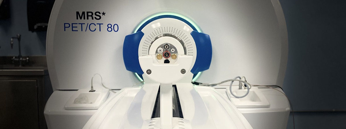
HSC teamed up with MR Solutions, a leader in preclinical imaging systems, to build the state-of-the-art facility. Our lab includes:
- a 7T Preclinical MR system based on dry magnet technology
- a floor-standing high-resolution CT system
- a PET system using clip-on technology to move between the CT and the MR systems
- and a multi-pinhole SPECT system based on the clip-on technology
Social media