Pre-clinical Imaging
Pre-clinical Imaging Core provides investigators with access to state-of-the-art multi-modality MRI/PET/CT/SPECT imaging technologies enabling researchers to image live animals on the organ, tissue, cell, or molecular level for basic and/or translational research. The Core is equipped with supporting and monitoring instrumentation to allow a wide variety of imaging studies.
Contact
Contact us for more details about equipment, services, and scheduling time in the Pre-clinical Imaging Core.
iLab
Log in to iLab to see a full list of equipment and schedule time in the Pre-clinical Imaging Core.
Per Hour Charges Based on User Type
| Internal | NEXT Lab | External | ||
| MRI | $ 125.00 | $ 175.00 | $ 350.00 | |
| PET/MRI |
$ 175.00 | $ 245.00 | $ 490.00 | |
| CT | $ 50.00 | $ 70.00 | $ 140.00 | |
| PET/SPECT | $ 75.00 | $ 105.00 | $ 210.00 | |
| PET/CT | $ 100.00 | $ 140.00 | $ 280.00 |
Authorized Experienced Internal Users
| Off Hours | Regular Block | Off Block | |
| MRI | $ 85.00 | $2100 / 24 hrs | $1750 / 24 hrs |
| CT | $ 40.00 | $350 / 8 hrs | $270 / 8 hrs |
- MR Solutions MRI scanner with Variable Fields at 7T and 3T
- MR Solutions sequential PET/CT and PET/MR scanner
- MR Solutions sequential SPECT/CT and/or SPECT/MR imaging
- Animal physiology monitoring equipment
- Perkin-Elmer IVIS Imaging System
- Image-analysis station for facility users
- Refrigerators and freezers
- Gamma counters
- Short-term animal housing for radioactive material (RAM) decay
Pre-clinical imaging strategies in numerous applications includes:
- Non-invasive quantitative assessment of disease progression
- Physiologic, functional and metabolic imaging
- Small molecules development
Instrumentation Manuals from MR Solutions
Starting from May 15, 2022, participants will be required to complete the online training before entering the Imaging Center. The training will certify that students have reviewed and completed the Radiation and MRI Level 1 Training.
Safety Training for Imaging Core is a self-paced training required for working in environments where radiation is utilized for the first time as per the HSC Distance Education Policy. Please read the following steps to get started:
- Click on the “Objectives” tab on the left to get familiar with the course structure and assessments.
- Click on the “Modules” tab on the left and complete “Module 0: Getting Started.”
- Ask any course-related questions in the “Q&A Forum” or reach out to the course coordinator for further assistance.
Existing HSC students can click here and self-enroll in the course. Non-HSC students/employees (if they not able to enroll from the link) an account would be required to set up access
This course will provide you with a basic safety understanding of MRI and radiation management.
- Access Request
- Radiation Safety
- Training Request
- IACUC Procedures and Guidelines
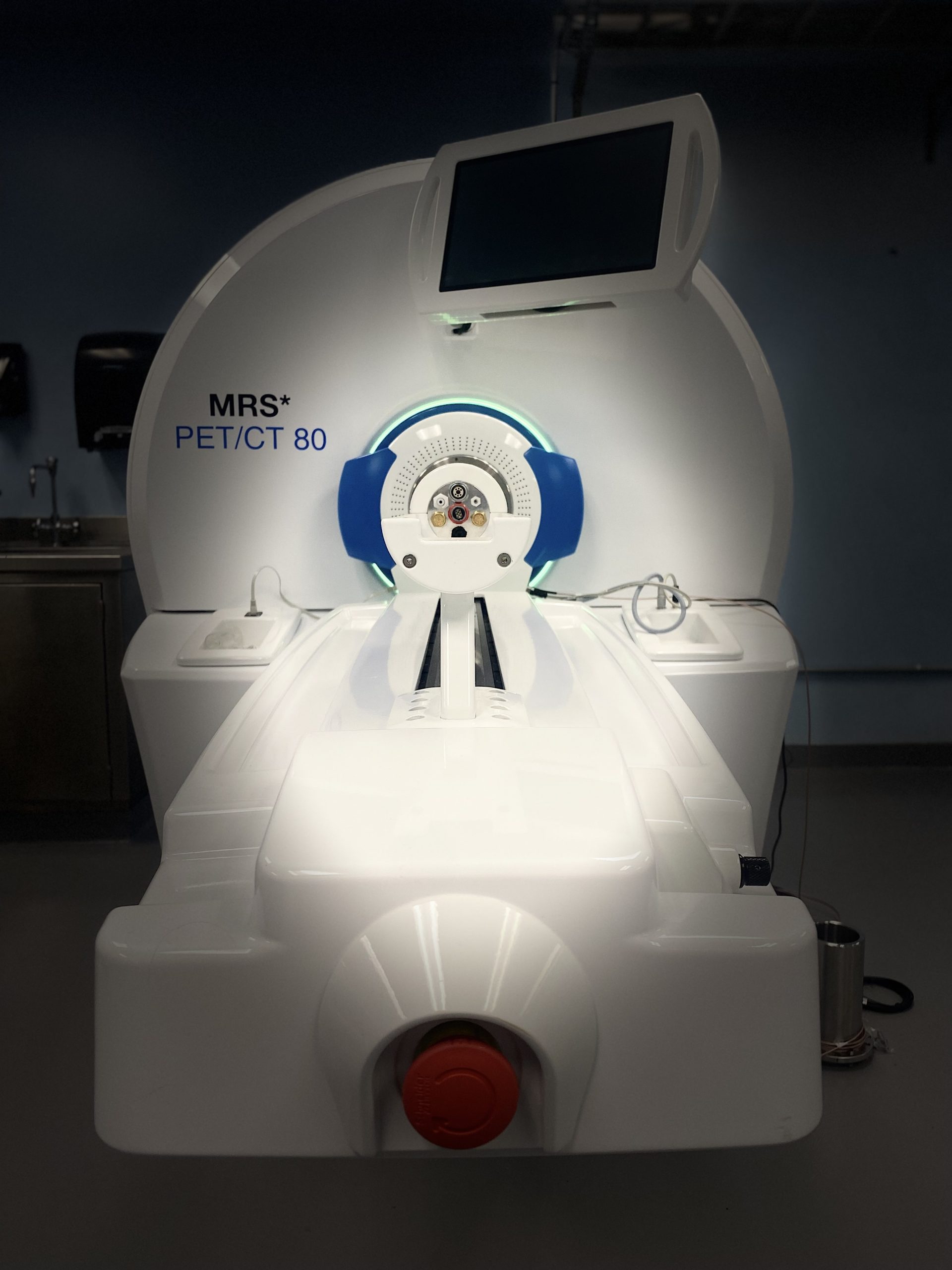
The Core currently has a state-of-the-art cryogen-free MRI magnet, MRS*DRYMAG7017, with a variable field capability to change between 3T and 7T fields with a 17-cm bore magnet equipped with a 600 mT/m RF gradient that will enable high-field imaging studies (i.e., EPI, DTI, DWI, spectroscopy) at 7T while delivering direct clinical translational capabilities at 3T. RF coils are available for different size subjects (mice and rats) and both field strengths. PET scanner (MRS*PET-CO 802) can be coupled with the MRI gantry or the CT gantry for optimized multimodality imaging with Tansaxial FOV of 80.0 mm, Axial FOV of 102.48 mm, and Spatial Resolution of 0.7 mm. SPECT CLIP-ON for Sequential SPECT/CT and SPECT/MR imaging and a high-resolution CT scanner (MRS*CT80). The modular design of the scanners enables the user to perform stand-alone MRI, PET, or CT imaging, or the systems can be coupled together in seconds to perform sequential PET/MRI or PET/CT imaging, optimizing performance and workflow.
Contributions to scientific output is an important metric for Core Labs, as it enables us to obtain financial and other support to ensure continuous high standards of operations to foster innovative research.
Users are encouraged to acknowledge Pre-clinical Imaging Core in presentations, posters, papers, and all other publications.
For example: “We acknowledge [name of staff] in the HSC Pre-clinical Imaging Core for training and assistance on [following]”.
When to acknowledge or provide co-authorship
- Include an acknowledgement any time the staff member or the Core provides services that support your research
- If a staff member has made a significant intellectual contribution beyond routine sample analysis, please consider co-authorship