Select Category:

Image taken in Microscopy Core featured on Journal of Neurophysiology cover page

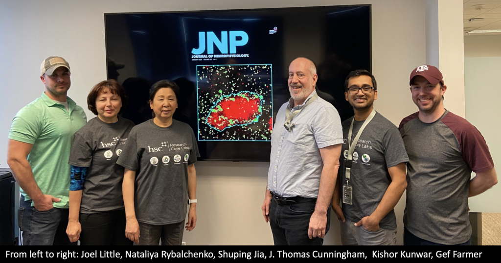
We are thrilled to find out confocal image taken in the Microscopy Core by Shuping Jia from the Cunningham lab and Kishor Kunwar from the Microscopy Core is being featured on the cover of the Journal of Neurophysiology. This is a remarkable achievement and a testament to these researchers' hard work and dedication.

Continue reading "Image taken in Microscopy Core featured on Journal of Neurophysiology cover page"

Genomics Core manager update

Taegun Kwon headshot

Dr. Taegun Kwon is joining the Genomics Core as its new manager starting Jan 17th, 2023. With a Ph.D. from The University of Texas at Austin and over 7 years of experience in the genomics field, Dr. Kwon brings a wealth of expertise and experience to the role. He is excited to be working closely with researchers in and around HSC to understand their needs and ensure that the Core is able to provide the necessary resources and expertise to support the innovative work.

Continue reading "Genomics Core manager update"

Genomics Core acquiring new single-cell technologies

Genomics Core is excited to announce the upcoming arrival of Chromium X Series and Visium CytAssist from 10x genomics. These cutting-edge platforms will provide researchers with powerful new tools for single-cell genomics and spatial transcriptomics research.

Continue reading "Genomics Core acquiring new single-cell technologies"

Microscopy Core adds Huygens-Image processing and analysis software to its toolkit

Huygens Essential is a powerful imaging software that allows users to process and analyze large sets of microscopy data. Developed by Scientific Volume Imaging, it is compatible with a wide range of microscope platforms, including widefield, confocal, and super-resolution systems. Huygens Essential offers a wide range of advanced image processing tools, including deconvolution, registration, and segmentation. It also provides support for a variety of file formats, including image stacks, multi-dimensional datasets, and time-lapse sequences.

Continue reading "Microscopy Core adds Huygens-Image processing and analysis software to its toolkit"

Opening Soon – Histology Core

A new Histology Core Lab is set to open by end of January 2023, providing HSC researchers and external clients with a state-of-the-art resource for processing and analyzing tissue samples. The Core will be equipped with the latest technology, including an automated slide scanner, tissue processor, and a variety of staining and imaging techniques.

Continue reading "Opening Soon – Histology Core"