School of Biomedical Sciences

Connectomics
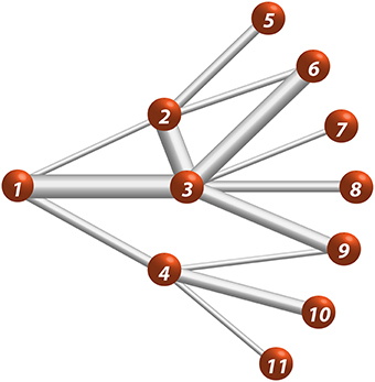
Connectomics refers to the study of neuroscience concerned with connectivity principles of the brain network. The brain network is the interconneconnected physical system of principal constituents and interactions between them. Traditionally we refer to three types of connectomes; macroscale, mesoscale and microscale. Macroscale connectomics refers to large scale connectivity using MRI (diffusion and fMRI) or any other methodology in which the basic constituent in the order of brain regions or individual scalp electrodes etc. The microscale refers to the mapping where the basic constituent is individual neurons and all its synapses. The mesoscale refers to in between scale between the macro and microscales, where the basic constituents are small regions; like cortical columns or specific neuronal types within brain regions. In the lab, we are interested in the macroscale study of brain network to identify translational biomarkers of brain health that we may translate from animal studies to human or viceversa
TVFC
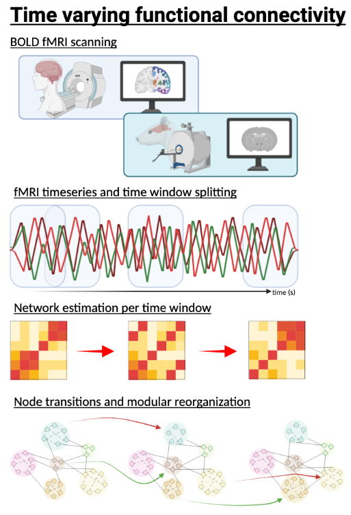
Time-varying functional connectivity is any method to estimate time-resolved fluctuations in the brain’s functional connectivity. The brain is a complex system in its physical connectivity and temporal relationships. Neuroimaging data is primed for this endeavor, as it provides us with a comprehensive dataset to identify the spatiotemporal principles of brain function. These features are of utmost importance to gain new insights relating the brain connectivity to cognition in health and disease. In the lab, we seek to apply these methods and develop new ways of obtaining time-resolved brain features. To do this, we mainly employ graph-theoretical principles of network dynamics to model the brain’s dynamics role in behavior.
Substance use Disorders

Substance Use Disorders is a mental disorder that affects a person’s ability to control their use of drug or medication. This disorder start with the sporadic use of a rewarding substance and as time progress, users develop tolerance and need larger amounts of the drug to obtain the rewarding effects of it. With increased drug use, the brain responds with changes in its structure and function. In the lab we are interested in elucidating the important features of this disorder using neuroimaging, and novel neuroscience techniques (e.g., optogenetics, self-administration, cognitive testing). We hope to identify important insights that can lead to novel interventions and therapies to alleviate the high financial and societal cost of these disorders.
Neuroimaging
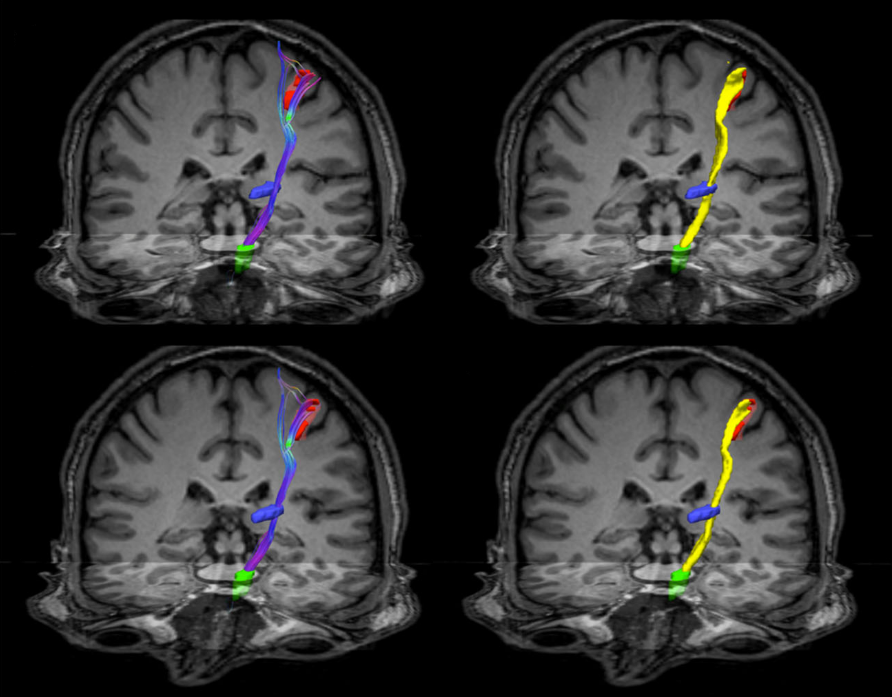
Neuroimaging is the process of generating images of the brain and is a powerful approach to gain a glimpse of the inner workings of the brain and its structure. Neuroimaging can refer to any methodology to captures pictures of brain; examples are MRI, CT scans, PET scans, fNIRS, among others. All provide information about a distinct feature of brain structure, function, or metabolism. In the lab, we focus mainly on MRI studies to gain insights about the function and structure of the brain. We employ diffusion MRI to generate maps of connectivity or of the microstructure of the brain. We also employ fMRI techniques to gain insights regarding the organizational principles of brain activity and dynamics. This toolbox allow us to gain a noninvasive view of the brain and obtain important information about the brain’s functional and structural reconfiguration in animal models of neuropsychiatric disorders and human subjects.
Social media