Light Microscopy Resources
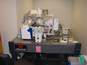
Zeiss LSM 510
Meta-head – for normal confocal including a spectrophotometric linear ccd array
Live-scan head – for capturing short duration events (i.e. Calcium Sparks). Uses line-scanning rather than point scanning (not as high a resolution image as with the Meta-head)
Incubator Enclosure – Maintains user set CO2 level, temperature & humidity around the sample stage & objectives. For live cell experiments.
Installed Lasers:
Diode 405nm -50W
Argon/2 561nm
Diode 488nm -100W
DPSS 532nm -75W
DPSS 561nm -10W
HeNe 633nm
Diode 635nm -35W
Objectives:
10x, 0.45NA, 2 mm WD (air DIC), Plan-Apochromat
20x, 0.8NA, 0.55 mm WD (air DIC), Plan-Apochromat
63x, 1.4NA, 0.19 mm WD (oil DIC),Plan-Apochromat
40x, 1.2NA, 0.28 WD (water), C-Apochromat
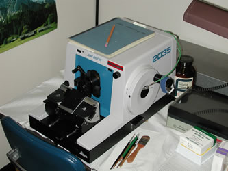
Leica Biocut
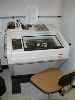
Leica Frigocut Cryostat
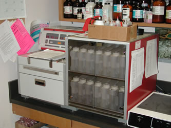
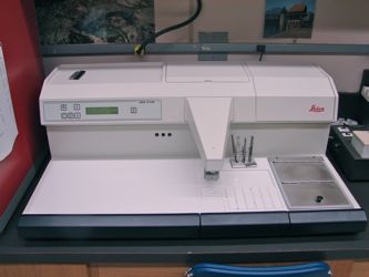
Histo-Tissue Processor
Paraffin Embedding Station
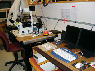
Olympus TIRF (Total Internal Reflectance Fluorescence) Microscope
Able to image in real time cell membrane fluorescent labels (i.e. GFP)
- may be able to use for FRET as well (currently researching this possibility)
- Also can be used for calcium imaging
Social media