|
|
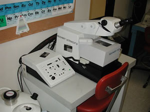
Leica Ultracut 5 Ultramicrotome/Cryoultramicrotome
Used to cut ultra-thin sections for transmission electron microscopy
- Plastic sections
- Frozen (cryo) sections
Can use glass, diamond or sapphire knives. |
 |
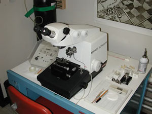
Leica Ultracut 5 Ultramicrotome
Used to cut ultra-thin sections for transmission electron microscopy
Can use glass, diamond or sapphire knives. |
 |
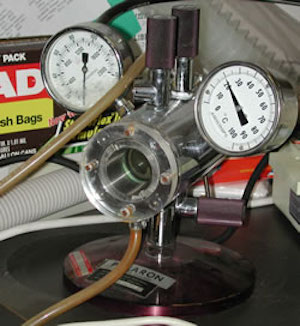
Polaron Critical Point Dryer
Used to dry biological samples for observation on a scanning electron microscope
- Avoids the formation of a liquid/gas interface during the drying process
- Organic solvent (i.e. EtOH or Acetone) is substituted with a liquified gas (CO2) within the sealed chamber.
- The temperature and pressure are then raised above the triple point for CO2 at which point the liquid CO2 transitions from a liquid to a gas throughout the chamber and the sample without the formation of a liquid/gas boundary.
- The gas is then vented and the dried sample removed.
|
 |
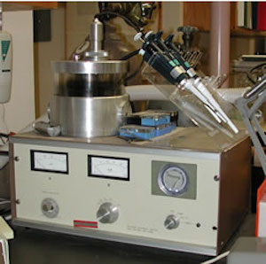
Polaron Sputter Coater
For evaporation of a continuous thin conductive film over a sample destined for the scanning electron microscope
- Uses a gold/palladium target for the coating.
- Uses the noble gas Argon to produce a non-oxidizing poor vacuum to sputter coat through.
|
 |
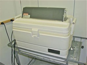
Low-Temp UV Polymerization Chamber
Used to polymerize immunocytochemistry samples which have been infiltrated with resins that helps to maximize the antigenicity of the antigens which will be probed for.
- uses dry ice (solid CO2) to maintain a low temperature within the chamber
- uses an ultraviolet (UV) lamp to polymerize the resin
- mostly use acrylic and methacrylic resins formulated to polymerize with UV light
- LR White
- LR Gold
- Lowicryl
|
 |
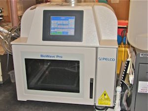
Pelco BioWave Pro Microwave Processor
Uses microwaves to speed up the protocols used to prepare samples for the microscope
- does not use heat (not cooking the samples)
- uses the increased molecular motion provided by the microwaves to speed up fluid exchanges and molecular reactions (i.e. fixation)
- Used to process biological samples for electron microscopy and light microscopy
- Used to stain sections for the TEM and light microscopy.
|
 |
Social media